ScienceHunter 11.07.2019
Automatic Processing and Analysis of Digital Images of Different Types of Human Cells
The analysis of biomedical images has long become an integral component, both in diagnosing diseases and as applied to therapeutic purposes. This is due to the fact that a fairly large proportion of the total volume of all data in medicine is images. It is an important source of patient health data.
Biomedical imaging technologies use a wide range of different opportunities such as X-rays, magnetism (MRI), computed tomography scan (CT scan), SPECT, OCT, ultrasound, endoscopy, etc.
Since recently, an analysis of digital images of biological fluids, primarily, blood cells, cerebrospinal fluid, buccal epithelium scrapings and others, has been added to this list. Examples of such images are presented in Fig. 1.
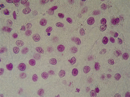
Fig. 1. Scan of nuclei scraping of buccal epithelial cells
A biological fluid swab often provides the primary and, sometimes, the only evidence of a specific diagnosis, remaining an important diagnostic tool even in the molecular analysis era of molecular analysis with the ability to isolate DNA, RNA and other material in the cell structure. An interest in this topic is driven, first of all, by an opportunity to automate the process of diagnostics, which is reduced to analysis of digital images of the respective human cells.
At the present time, there are few automated tools that would help doctors fulfill the routine duties of analyzing images, helping to process and analyze such information. Most of these systems are built on the use of neural network data processing technologies (artificial intelligence) with their inherent disadvantages - the need for training on a very large amount of data and the inability to interpret the results.
Our approach is based on the extraction of various features of cells and the formation of a training sample from it for subsequent classification, provides visibility of the results and shows high accuracy on test data. The system was developed on the basis of the work results that are provided by our specialists in the field of image processing. As a result, we built a system, which is able to segment the images of cells and extract the necessary features from them to build an object-feature table (Fig. 2). A brief presentation is available here.
Direct work with the web service for the automatic processing and analysis of digital images of human cells is possible on the site.
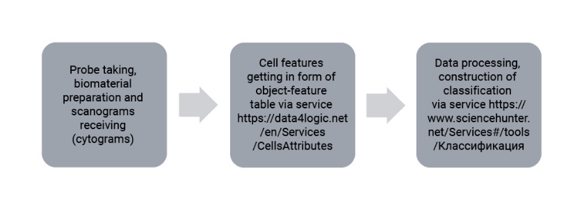
Fig. 2. The scheme of service
When analyzing any image, it is divided into objects and background. Therefore, the primary goal of image analysis is a process of segmentation, aimed at identification of the areas, corresponding to the objects, and relation of the remaining part of the image to the background. Success in image analyzing depends, primarily, on reliability and correctness of segmentation, which is quite a difficult task. Its solution allowed us to proceed to the construction of a recommender system for the pre-processing of cell images. The result of the automatic analysis system is shown in Fig. 3.
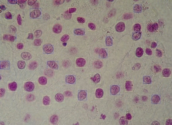
Fig. 3. Image scraping of buccal epithelium after treatment
The developed software is designed to improve the efficiency of medical personnel, (including research), in the diagnosis, treatment and monitoring of various diseases that have manifestations at the cellular level. Moreover, in our opinion, further integration of a recommendation system and a computer diagnostic system built on the basis of neural networks will provide an automated medical image processing system for subsequent early non-invasive diagnosis of breast cancer, and possibly other types of cancer.
Furthermore, one can expect to expand the spectrum of use of the system to analyze not only the fluid of mucous membrane, but also, for example, lymphocytes, eosinophils, biopsy samples of lesions of the brain, teeth, other body systems and other types of human cells.
So far, the system functions assume analysis of 2d images (3d is not supported), however, the fundamental algorithms already show fairly accurate results, helping professionals quickly track the increase in the number of a certain cell type on the sample, see their structure, and, therefore, more accurately diagnose the processes occurring in human organs, see signs of various diseases at an early stage.
And, finally, we are open for cooperation both with people from science and with commercial organizations of any kind.